Brain Tumor Detection Accelerated to Two Hours in Operating Room
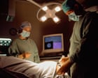
Revolutionary Brain Tumor Diagnosis Technology Transforms Patient Care
A groundbreaking brain tumor diagnosis system has emerged as a transformative tool in modern neurosurgery, enabling medical teams to identify and classify tumors within two hours while patients remain on the operating table. This innovative approach represents a significant departure from traditional diagnostic methods, which typically required eight weeks or longer to complete comprehensive analysis.
How the Intraoperative Testing Works
The intraoperative tumor testing methodology operates by analyzing tissue samples obtained during surgery in real-time, allowing surgeons to receive critical diagnostic information without delays. NHS England has recognized the potential of this technology and has initiated a pilot program across five specialized centers nationwide. The system enables medical professionals to make informed decisions about treatment protocols and surgical approaches while the patient is still under anesthesia, eliminating the need for additional procedures and repeated imaging sessions.
Clinical Benefits and Patient Impact
Medical experts have characterized this development as a significant advancement, particularly for patients whose conditions demand immediate intervention. The rapid brain tumor detection capability proves invaluable for individuals who, as physicians note, "do not have time on their side." Early and accurate diagnosis directly correlates with improved treatment outcomes, increased survival rates, and enhanced quality of life for affected patients. By compressing the diagnostic window from weeks to hours, clinicians can initiate targeted therapeutic strategies without unnecessary delays.
Operational Implementation in Surgery
The technology operates seamlessly within the surgical environment, requiring no additional patient transfers or extended hospital stays specifically for diagnostic purposes. Teams can perform surgical tumor identification immediately upon tissue extraction, with results available before the operating team completes the initial surgical intervention. This integration streamlines the entire care pathway, reducing patient anxiety and accelerating the transition to definitive treatment plans.
Pilot Program Expansion Across NHS Centers
Five designated centers across England are currently evaluating this NHS diagnostic innovation through structured pilot initiatives. These facilities represent diverse patient populations and surgical environments, providing comprehensive data on system performance, reliability, and clinical applicability. The pilot phase aims to establish standardized protocols, train surgical teams, and document outcomes that will inform broader implementation strategies across the National Health Service.
Future Prospects and Medical Advancement
The successful deployment of this diagnostic method promises to reshape how medical institutions approach brain tumor management. Scientists and healthcare administrators anticipate that widespread adoption could establish new standards of care, potentially reducing unnecessary diagnostic delays that currently impact thousands of patients annually. The combination of accuracy, speed, and real-time surgical integration positions this technology as a cornerstone of next-generation neurosurgical practice, potentially inspiring similar innovations across other medical specialties requiring rapid tissue analysis and classification.